Explaining 3D Scans at the Periodontist's Office
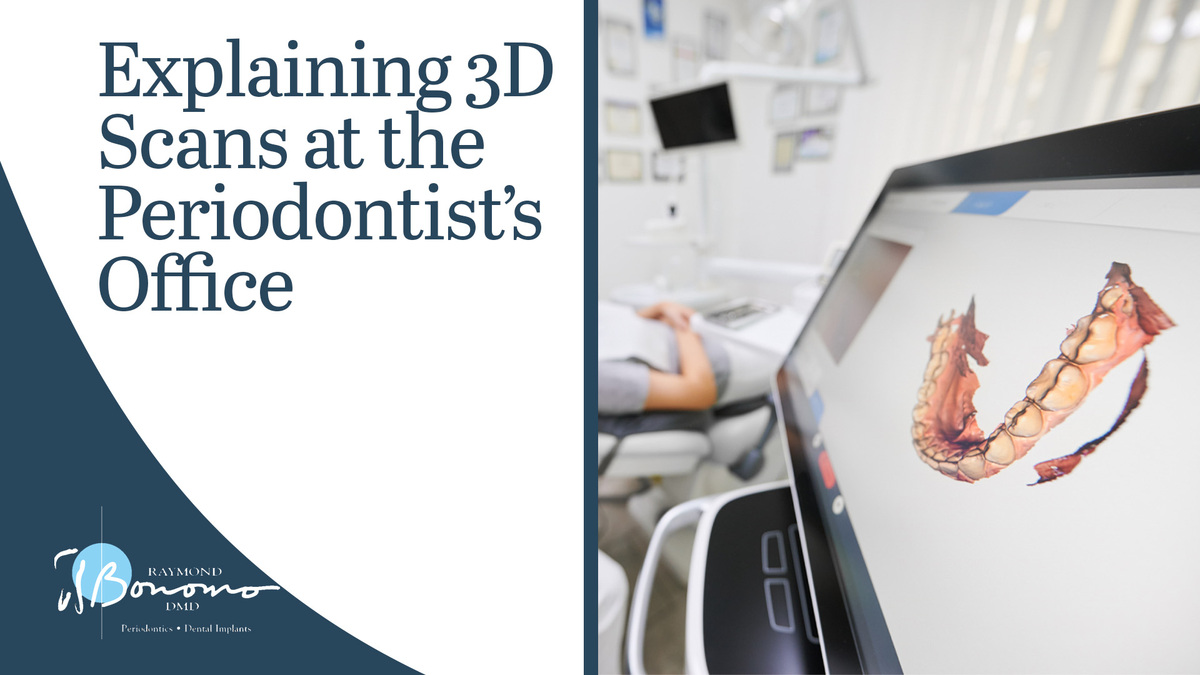
If you are visiting the periodontist about gum or oral health concerns, then your doctor may recommend that you undergo a 3D scan or 3D image procedure so they can get a better understanding of the internal structure of your soft tissues and bones. These images help your periodontist with treatment planning for procedures like dental implants.
Many patients have questions about what the process of 3D scanning or 3D imaging looks like for their dental structure. They often wonder if they will be x-rayed or exposed to radiation, how long the process will take, what their periodontist will do with these detailed images, what dental procedures 3D scanning is right for, and more. These are all perfectly valid questions, and patients should know these answers before undergoing a 3D imaging procedure.
In this post, we will answer your most pressing questions about 3D scans at the periodontist's office. We will discuss the process of 3D scanning and what it accomplishes. In addition, we will discuss some frequently asked questions about the process of 3D imaging for periodontal care. Keep reading to learn more!
Introduction to 3D Imaging in Periodontics
3D imaging has revolutionized the field of periodontics, providing clinicians with a more detailed and comprehensive view of the oral cavity. From diagnosing periodontal disease to planning for dental implants, 3D imaging offers a level of precision and accuracy that was not previously possible with traditional two-dimensional radiographs. This advanced technology allows for better treatment planning and more predictable outcomes in periodontal therapy.
In this article, we will explore the different types of 3D imaging used in periodontics, the benefits it offers for both clinicians and patients, and the implications for the future of periodontal care.
The Process of 3D Scanning
3D scanning involves the process of capturing digital images of an object's surface to create a 3D model. The first step is to position the object on the scanning platform. Then, a 3D scanner is used to capture images of the object from multiple angles. These images are then processed by specialized software to create a 3D model.
Resolution is crucial in 3D scanning, as it determines the level of detail and accuracy of the captured images. High resolution is essential in all three planes (x, y, and z) to ensure that the 3D model accurately represents the object. This is particularly important for clinicians when scanning prepared teeth for dental applications. Higher resolution enables the accurate capture of fine details, such as occlusal morphology and margin precision, which is crucial for creating dental restorations that fit precisely.
3D scanning involves capturing digital images of an object's surface from multiple angles, with resolution being essential in all three planes to ensure accuracy. For clinicians scanning prepared teeth, higher resolution is especially vital for capturing the minute details necessary for creating precise dental restorations.
Frequently Asked Questions (FAQs)
Let’s look at some of the most frequent questions patients have about 3D imaging.
What are Cone Beam and 3D Imaging?
Cone Beam and 3D Imaging are advanced imaging technologies that provide detailed images of the entire craniofacial structure, including teeth, bones, and connective tissues.
How can Cone Beam and 3D Imaging benefit patients?
These technologies offer a comprehensive view of the oral and maxillofacial regions, allowing for a more accurate diagnosis and treatment planning. These detailed images help in identifying any abnormalities or potential issues that may not be visible with traditional 2D imaging. This leads to more precise and tailored treatment plans for patients.
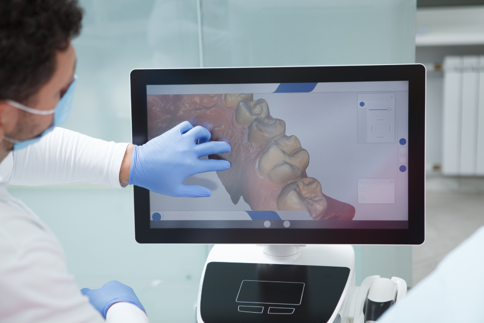
How do these images help in oral surgery and dental treatment planning?
The 3D images generated by Cone Beam and 3D Imaging can be dissected and manipulated to assess the exact dimensions, angles, and relationships of the structures within the oral cavity. This level of precision ensures that oral surgeries, such as dental implants or orthognathic surgery, can be planned with accuracy, leading to better outcomes for patients.
3D Scanning at Bonomo Periodontics
3D scanning is common when pursuing periodontic treatment. A completely painless procedure, 3D imaging gives your doctor a better understanding of the internal structure of your mouth, bones, and sinus cavities using cutting-edge technology, and superior image quality.
This single scan procedure is highly effective at helping your periodontist identify oral issues and make an informed diagnosis. Unlike two-dimensional images, three-dimensional images produced by this process allow your doctor to get a more comprehensive understanding of your periodontics needs.
If you are interested in seeking periodontal treatment or have questions about dental implants, Bonomo Periodontics is here for you. We are Cincinnati's most trusted source of periodontal care, and Dr. Raymond Bonomo is renowned for his ability to treat gum disease with care.
Reach out to our office about your periodontal needs by clicking here!
Follow Bonomo Periodontics on Facebook for more information!
Happy with the difference Bonomo Periodontics has made in your life? Leave us a five-star review here!